BIO
135 Virtual Labs
Epithelial
Tissues Identification Guide
Return to:
Main Tissues Page ![]() BIO 135 Main Page
BIO 135 Main Page ![]() Lab 3 Page
Lab 3 Page
![]()
|
|
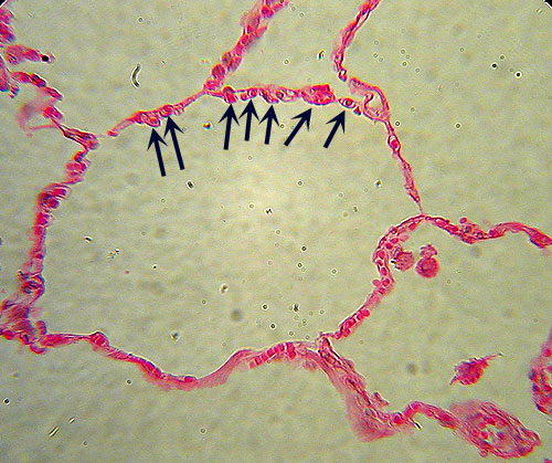
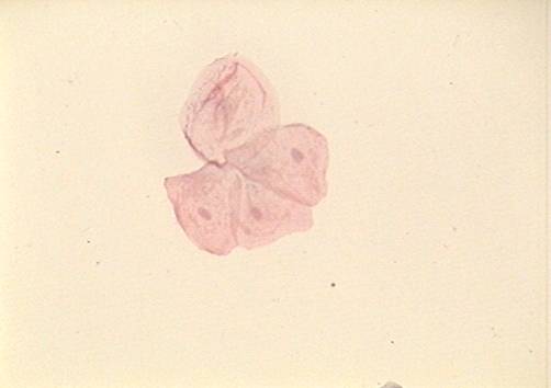
Simple Squamous Epithelium
Identification: Small, flat cells arranged around
large, empty circles (air sacs). May be confused with Adipose Tissue, but note the multiple
cells and nuclei (arrows). Features
to Know: nuclei. Where
Located: lung (air sacs or alveoli) and lining blood vessels . Function: diffusion
(gas exchange). The
top image is what you would see with a transverse section through the
tissues. The bottom image is from a
cheek smear and shows a cluster of four cells viewed if they were laid out
flat. |
|
|
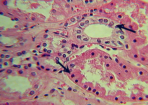
Simple Cuboidal Epithelium
Identification: Squarish cells with round nuclei
in a single row (arrows), usually arranged in a circle (tubule). Features
to Know: nuclei. Where
Located: kidney tubules (can also be seen in sweat glands of skin slide). Function: absorption
and secretion. |
|
|
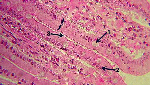
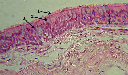
Simple Columnar Epithelium
Identification: Tall rectangular cells, with single,
neat row of oval nuclei, usually more towards the base (in the bracketed
row, the base is towards the top), leaving an apical region of nucleus-free
cytoplasm. Note also goblet cells, found only here and in Pseudostratified
Columnar Epithelium. Features
to Know: goblet cells (1), nuclei (2), microvilli (3). Where
Located: jejunum of small intestine. Function: absorption
and secretion. |
|
|
Pseudostratified Columnar Epithelium
Identification: Tall rectangular cells, with multiple
irregular rows of nuclei (bracketed; compare to simple columnar, above). Note
also goblet cells, found only here and in Simple Columnar Epithelium. Features
to Know: cilia (1), goblet cells (2), nuclei (3). Where
Located: trachea. Function: secretion
and movement of mucus. |
|
|
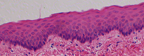
Stratified Squamous Epithelium
Identification: Many layers (6 or more) of small,
flattened cells. The only other epithelial tissue with so many layers is
transitional (below), but note that stratified squamous epithelium typically
has a more evenly contoured surface; with the uppermost layers of cells
flattened. Features
to Know: nuclei, if evident. Where
Located: lining mouth and esophagus. Function: protection
from abrasion and infection. |
|
|
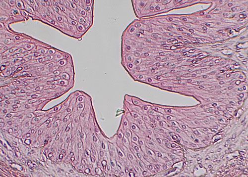
Transitional Epithelium
Identification: Numerous layers of cells of varying
and often irregular shape, though generally not squamous (when unstretched as
in the slides). Surface of tissue is folded (inside of ureter) or bumpy
appearing (urinary bladder). Features
to Know: nuclei. Where
Located: ureter (and urinary bladder). Function: elasticity:
stretch and retract. |
You will
not need to know either stratified cuboidal or stratified columnar epithelia in
lab.
![]()
Continue
on to the Muscle & Nervous Tissues Page
Continue on to the Connective Tissue Page
![]()
This page created by Udo M.
Savalli. Maintained by Bill D.
Snyder Last updated September 23, 2009